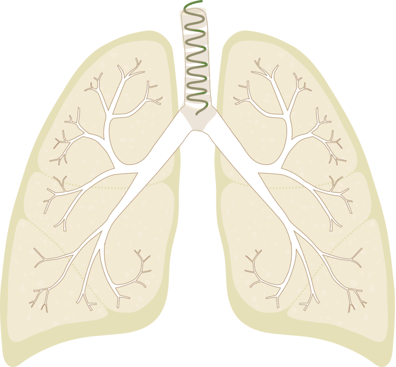
FORCED OSCILLATION TECHNIQUE
Forced Oscillation Technique (FOT) is a noninvasive method to assess lung function during normal breathing. Differently from spirometry, where the subject is requested to forcefully and repeatedly exhale all the air from the maximum to the lowest reachable lung volume, FOT uses a small pressure oscillation (equivalent to the pressure exerted by 2 cm of water) over imposed on regular quiet breathing to “force” a small quantity of air to move in and out the respiratory system. To separate the response of the respiratory system from the spontaneous breathing activity, the respiratory system is stimulated using pressure oscillations that are at least 10 times faster of the human breathing frequencies ranging from 4 Hz up to 40 Hz. Since slow pressures (i.e. < 10 Hz) are more able to probe lung periphery ((Bates & Suki, 2008) they are used to measure the entire system alone or in combination with faster stimuli.
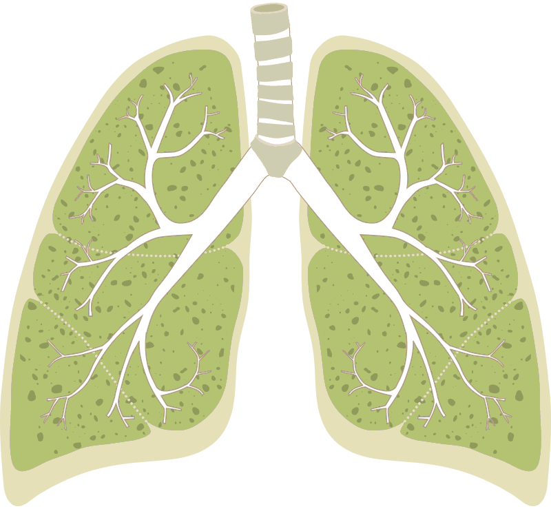
What it measures
A FOT test measures how easy is for the air to move in and out the respiratory system. Such “impediment” is quantified by combining the force used to move the air (i.e. the pressure) with the amount of air that actually has been displaced (i.e. air flow) and calculating the RESPIRATORY IMPEDANCE. Air can be prevented to move in and out the respiratory system by several factors. Airway obstructions, reduced lung parenchyma elasticity, peripheral obstruction or alveolar collapse are all elements that will produce an increase of the RESPIRATORY IMPEDANCE.
Mathematically it is possible to separate such contributions by splitting IMPEDANCE in into RESPIRATORY RESISTANCE (Rrs) and REACTANCE (Xrs).
Rrs – Respiratory Resistance
For each respiratory segment, resistance is a measurement of its degree of obstruction. The tracheobronchial tree is a complex branching structure of respiratory segments and respiratory resistance measured by FOT is the result of the combination of the resistances of all the stimulated segments. This complex structure makes Resistance very sensitive to the degree of obstruction of the main central airways while small airways (< 2 mm in diameter) account for only 10% of total airway resistance ((Mead, 1970)). Resistance of healthy adults subjects is almost independent by the frequency of the stimulating pressure and it became abnormally higher at lower frequencies during severe obstruction (i.e. COPD GOLD stage 3-4(Dellacà, Pompilio, et al., 2009) or during positive bronchial challenge(van Noord, Clement, van de Woestijne, & Demedts, 1989)

Xrs – Respiratory Reactance
At lower stimulating frequencies, respiratory reactance is a measure of the elastic recoil forces of the respiratory system. It enables clinicians to determine how effectively the lung is being ventilated in its distal areas or how well air reaches the peripheral areas (Dellacà, Andersson Olerud, et al., 2009). Xrs falls below predicted values at low oscillating frequencies in the following conditions:
-
-
- peripheral obstruction(Downie et al., 2013)
- presence of tidal expiratory flow limitation (Dellacà et al., 2007)
- alveolar gas trapping and/or closing of alveolar units (Dellacà, Andersson Olerud, et al., 2009)
-
Xrs can be also affected by chest wall restrictions (i.e. obesity, kyphoscoliosis, pregnancy, etc)
RESTECH INNOVATION
Respiratory system is a very complex active structure made up by soft and hard tissues that move and interact during the breathing activity. During inspiration, respiratory muscles expand the rib cage to reduce the alveolar pressure and initiate a flow of fresh air from the mouth/nose trough the larynx, vocal cord and airways down to the alveoli. During the expiration, the elastic recoil of the lung and rib cage (and, during exercise, the expiratory muscular activity) “squeeze” the air from the alveolar region along the airways, vocal cord, larynx to be expelled through the mouth/nose. Diseases that alter these, dynamic interactions produce specific within breath changes in the mechanical properties of the lung. Differently from spirometry and body plethysmography, FOT can measure respiratory function during normal breathing and, therefore, has the potential to measure within breath physiological and pathological changes in mechanical properties of the lung at its normally operating lung volume.

In last 15 years of academic and industrial research, RESTECH has developed innovative technologies that allow the assessment, in real time, of fast within-breath changes of Resistance and Reactance during the respiratory cycle that improve the diagnostic capability of the technique allowing:
- a more accurate assessment of the degree of the airway obstruction
- automatic detection of tidal expiratory flow limitation
- Accurate automatic on-line artifact removal
- Day-by-day remote monitoring of patients with chronic respiratory diseases with automatic detection of worsening periods
- Assessment of the degree of airways instability
Benefits
- Highly reproducible measurements made during tidal breathing
- No need to perform forced manoeuvres
- No need for supervision by specialist staff
- Test easy to perform by children, subjects with severe lung impairment or with neuromuscular diseases.
